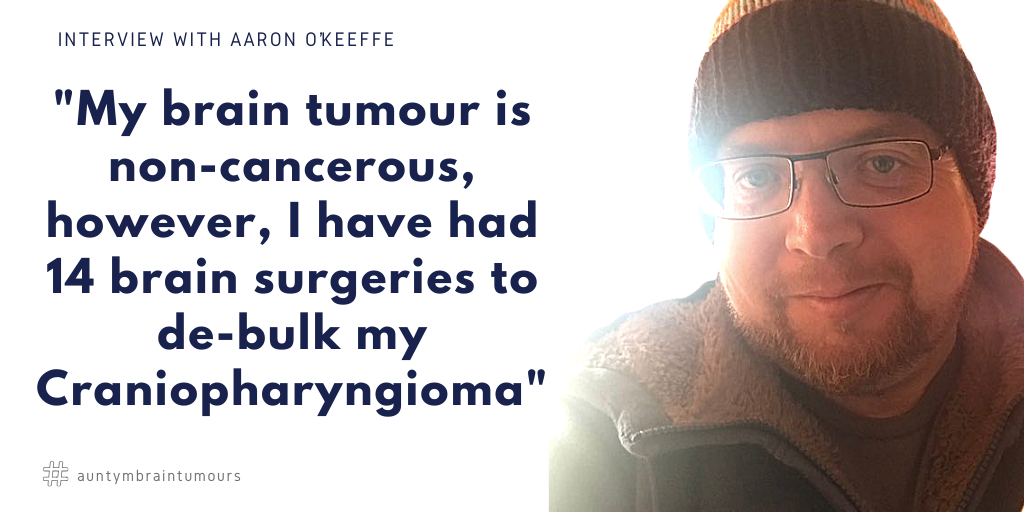
Living With a Craniopharyngioma That Keeps Coming Back
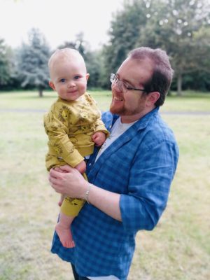
Aaron O’Keeffe, from Dublin, Ireland, has lived with a craniopharyngioma for more than three decades.
Since his diagnosis in 1991, Aaron has undergone 14 brain surgeries and two courses of radiotherapy. His tumour is non-cancerous, but its location near the pituitary gland and optic nerves has made it extremely difficult to treat.
Craniopharyngiomas are rare, usually benign brain tumours that often form near the pituitary gland and hypothalamus. Although they do not usually spread, their position can cause serious problems with vision, hormones, fatigue and quality of life. (Cancer.gov)
For Aaron, the hardest part has not only been surviving each operation.
It has been finding the strength to keep going.
A Diagnosis Hidden Behind Headaches
Aaron’s symptoms began in 1989, when he started having regular headaches.
Because his mother had a long history of migraines, the headaches were initially thought to be a family migraine problem. He was prescribed painkillers and sent home.
This continued for around two years.
Then, in November 1991, one of Aaron’s school friends suggested that he see another local doctor. That doctor also thought migraines were possible, but referred him for a CT scan just to be sure.
The scan changed everything.
It showed a brain tumour near Aaron’s pituitary gland.
Aaron now marks 12 November in his phone every year as “Craniopharyngioma Day” — the anniversary of the scan that found the tumour.
He does not remember much about the appointment itself, but he does remember being in the waiting room with his little sister Naomi. Together, they spotted a 20p coin under a vending machine and managed to retrieve it.
To them, it felt like a fortune.
They were excited about spending it on sweets.
But when they turned back to their parents, everything had changed.
When Vision Became the Warning Sign
Craniopharyngiomas can affect vision because they often grow close to the optic nerves and optic chiasm, the area where the two optic nerves cross. Symptoms can include headaches, visual problems and hormone changes. (Cancer Research UK)
For Aaron, visual changes became one of the clearest signs that the tumour was growing again.
After his diagnosis, his parents were told to watch closely for any changes. They made it through Christmas, but on 27 December, while watching television, Aaron realised he could not read the quiz show answers on the screen.
They were too blurred.
His parents contacted the hospital immediately.
The next day, Aaron was admitted for surgery.
He spent three weeks in hospital over New Year and into January. Because of where the tumour was, surgeons could not remove every tumour cell. His family were warned that recurrence was likely.
Aaron recovered well from the operation itself and was soon proudly showing visitors the 27 metal staples that had been removed from his healed incision.
Before long, he was back at school.
Life began to feel normal again.
But the tumour was not finished.

More Surgery, More Scars, More Uncertainty
Around 18 months later, in June 1993, a routine MRI scan showed new tumour growth.
Aaron had a second brain surgery in July.
Again, the tumour’s location made complete removal difficult. Because it was so close to the pituitary gland and surrounding brain structures, the surgical team had to balance removing as much tumour as possible with protecting Aaron’s brain function.
This time, to reduce the chance of regrowth, the team decided to remove Aaron’s pituitary gland completely.
Then, in December 1995, another MRI scan showed further tumour growth.
Aaron went back into hospital for his third brain surgery.
The surgeons reopened the scar on the right side of his head, but scar tissue made the operation difficult. Aaron woke up on the children’s ward on Monday evening and was told he would be going back to theatre again on Wednesday.
This time, they opened the other side of his head.
The scar stretched from the middle of his forehead to just in front of his left ear.
He was discharged in time for Christmas, with radiotherapy planned for early 1996.
Radiotherapy as a Teenager
Aaron’s first course of radiotherapy began in late February 1996.
He was in his third year of secondary school.
Every weekday for six weeks, he left home as normal, went to school, then escaped classes around lunchtime to travel into Dublin city centre. There, he met his mum before they travelled by bus to another hospital for treatment.
Before the sessions began, a plaster mould was made of his head. From that, a transparent plastic mask was created to keep his head still during treatment.
When the radiotherapy finished, Aaron was given both the mask and the plaster mould. The mask lived in the attic for years. The mould was painted and put on a shelf in his room.
For a while, the treatment appeared to work.
MRI scans and visual field tests continued regularly. Eventually, the appointments became less frequent. The tumour — or what was left of it — seemed to be behaving.
Then, in 2001, new growth appeared.
Because the radiotherapy had slowed the tumour’s growth, surgery was not immediately urgent. Aaron even managed to delay the operation until after a close friend’s 21st birthday celebrations.
It was a small piece of control in a life where so much had been decided by scans, symptoms and surgery dates.

When Hospital Memories Begin to Blur
Over time, Aaron’s hospital memories have become harder to separate.
He explains that his memory has slowly worsened over the years. Surgeries, admissions and hospital visits often blur together. Details from one operation mix with another. Events from different years become difficult to place.
That in itself is part of the long-term reality of living with a recurring brain tumour.
It is not just the surgery.
It is the years of monitoring, medication, scans, fear, waiting, recovery, and trying to return to ordinary life in between.
The Ommaya Reservoir: Trying a Less Invasive Option
After further tumour regrowth, Aaron’s surgical team began looking for less invasive ways to manage it.
During another operation, they inserted an Ommaya reservoir.
This is a device placed under the skin, connected by a tube to a fluid-filled area. In Aaron’s case, the hope was that doctors might be able to drain fluid from the cystic part of the tumour using a needle, without needing to open his skull again.
It was not automatic like a shunt. It had to be drained manually.
If it worked, it could reduce the need for future major surgery.
But with Aaron’s tumour, there was always an “if”.
Later, when he became unwell and was vomiting, doctors tried to drain fluid through the reservoir but could not remove anything significant. They suspected the tube had become blocked.
Aaron went back to theatre.
A second Ommaya reservoir was inserted, this time leading to a smaller bubble on the top of his skull.
Over the years, this second reservoir has been used a few times. The drained fluid has varied from around 5ml to 35ml and is often yellow or golden, sometimes brown or black.
Aaron says the procedure is not painful beyond the initial needle, but it creates a strange sensation.
The closest description, he says, is like a small balloon slowly deflating inside your head.
A Different Surgical Approach
As the years went on, repeated craniotomies became more difficult because of scar tissue and the effects of previous surgeries.
Aaron’s neurosurgeon suggested a different approach: transsphenoidal surgery, accessing the tumour through the nose instead of reopening the old scars on his head.
The surgery went well, and recovery was much quicker than after a standard craniotomy.
But then Aaron developed a cerebrospinal fluid leak.
CSF — the fluid surrounding the brain and spinal cord — began leaking slowly through his nose. This carried a risk of serious infection because the barrier between his nose and the inside of his head was not properly sealed.
Aaron returned to hospital.
A repair was attempted, and a lumbar drain was inserted into his lower back to reduce pressure while the repair healed. He had to lie flat for days at a time, only sitting up briefly for meals.
Every five days, the drain was switched off to see whether the leak had stopped.
It had not.
For almost a month, the leaking continued. Another operation was being arranged.
Then, just before surgery, the leak finally stopped.
That hospital stay lasted nearly six weeks — Aaron’s longest single admission.
Infection, MRSA and the Loss of a Bone Flap
One of the most difficult periods came after another surgery, when Aaron was discharged home but felt something was not right.
His wound was weeping fluid. He had severe headaches. His head began to swell.
When he returned to hospital, he was told he had an MRSA infection in his head.
He needed surgery again.
During the operation, infected material was cleaned out. The “bone flap” — the piece of skull that had been removed and replaced during earlier surgeries — was left out because doctors were concerned the infection might be lingering on it.
The plan was to replace it later with a modern plastic composite.
For Aaron, this was another reminder that the tumour itself was only part of the story. The complications, infections and consequences of repeated treatment could be just as difficult.
A Second Course of Radiotherapy
When Aaron had radiotherapy in 1996, he was told it was something he could only have once.
Years later, advances in treatment meant his doctors felt a second course could be delivered more safely and precisely.
For Aaron and his team, this was preferable to more surgery if it could help control the tumour.
His local hospital now had its own oncology centre, so instead of travelling across Dublin every day, treatment was only a short drive from home.
This second course involved 25 sessions over five weeks.
Again, Aaron was fitted with a mask to keep him still during treatment. This time, it was made from a plastic-like mesh.
After treatment ended, he was given the mask to keep.
It has since been spray-painted, framed and mounted above the stairs in his house.

When the Infection Returned
In late May the following year, Aaron noticed a small red mark on his left temple.
At first, it looked like a spot. But it had no head and slowly grew bigger. It felt soft, like it was filled with liquid. Sometimes fluid leaked from it, running down the side of his face or appearing on his pillow.
Aaron sent photos to his neurosurgeon on WhatsApp.
He was asked to come straight to the hospital emergency department.
The operation was relatively minor compared with what Aaron had been through before. The surgeon removed what Aaron called “the bubble” and sent samples to microbiology to check for MRSA.
Aaron was placed on long-term antibiotics.
But when the mark returned after the antibiotics ended, it became clear that the infection had not fully gone.
His neurosurgeon suspected MRSA was persisting on the old plastic tube from Aaron’s first Ommaya reservoir, which was still inside his head.
Removing it would be risky.
But it might be the best chance of clearing the infection completely.
Aaron had his 14th brain surgery.
Thankfully, the surgeon managed to remove the entire tube, and scans and lab results suggested the infected material had been removed or cleaned out.
Everything seemed to be going in the right direction.
Then Aaron had a seizure.
The Day His Family Knew Something Was Wrong
Two days after surgery, Aaron’s mum texted him.
He did not reply.
His dad texted him too.
Again, no reply.
Aaron’s phone was normally glued to his hand, so this was unusual. The messages had been delivered, but not read.
His mum rang. No answer.
A close friend visited the hospital that morning and found Aaron asleep in a single room. She left a book and did not wake him.
But Aaron’s mum still felt uneasy.
She rang the nurses’ station and asked someone to check on him. Then she left work and went straight to the hospital.
When she found Aaron, he was sitting on the bed, shirtless, with the windows open. He said he felt nauseous and warm.
A nurse who knew Aaron and his family well came into the room. When she asked him a simple question, Aaron stared back and did not answer.
His mum asked another question.
Again, no answer.
Then Aaron started breathing strangely and making exaggerated hand gestures, repeatedly saying, “No, no, no.”
His mum thought he might be having a stroke.
Doctors were called. A nurse held up a biro and asked Aaron what it was.
“I don’t know,” he replied.
He was sedated, intubated and sent for scans.
It was not a stroke.
Aaron had suffered a seizure.
He was moved to neurosurgical intensive care for closer monitoring before eventually returning to a single room. It was another three weeks before he was discharged home.
Life Now: Fatigue and the Invisible Effects
By June 2020, Aaron says his main health problem was tiredness and post-surgery fatigue.
He describes it as feeling like someone has flicked a switch in his head.
His brain suddenly stops processing information.
When that happens, he has to apologise and leave for somewhere quieter.
Fatigue is one of the common long-term effects people may experience after a brain tumour or treatment. Other effects can include changes to memory, concentration, vision, mood, speech or movement. (nhs.uk)
For Aaron, the impact is not always visible from the outside.
But it is real.
The Toughest Challenge: Keeping Going
When asked what the toughest challenge is for a survivor, Aaron’s answer is simple.
“For me, it is to keep going.”
After a brain tumour diagnosis, surgery or radiotherapy, many people are expected to recover and return to life as it was before. But that can be a long and difficult road.
You may have to rebuild your strength.
Return to work or study.
Spend time with family and friends again.
Learn how your body and brain now work.
And somehow, keep your head up through all of it.
For Aaron, this has been especially hard because the tumour has continued to return. His health problems have been such a large part of his life that it can be difficult to see past them.
But he keeps going.
“This little tumour has taken a lot from me,” he says, “and I’m sure shaped my life in ways I cannot see, but you’ve got to keep your head up and keep pushing forward.”
His way of thinking has always been:
As bad as it has been, it could always be worse.
He is still here.
Two arms, two legs, and a brain that still works — at least most of the time.
The Importance of Family, Friends and Support
Aaron is clear that he could not have survived without his family and close friends.
They have been his crutch through diagnosis, surgeries, radiotherapy, infections, seizures and recovery.
Their support has carried him through the moments when keeping going felt hardest.
His story is also a reminder that no one should have to face a brain tumour diagnosis alone.
Support groups and charities can help people understand what to expect, ask better questions, prepare for treatment and connect with others who understand.
 |
 |
Aaron’s Advice for Others Facing a Daunting Diagnosis
Aaron’s first piece of advice is direct:
Keep your head up.
Find a way to stay positive, even if the bright side feels tiny.
He also encourages people to look for support groups. Whatever your diagnosis, there is often a community of people who understand the doctors, hospitals, treatments and recovery process you may be facing.
You do not have to wait until after treatment to contact them.
Aaron also has strong advice about searching online:
Stay away from Dr Google.
Online information can be overwhelming, and it is easy to focus only on the frightening parts. If there is a 2% chance of something bad happening, that may be the only thing your mind remembers.
Most of all, Aaron says:
Use the nurses.
In difficult appointments, especially when receiving bad news, it can be hard to absorb everything a doctor says. A nurse can help fill in the gaps afterwards, explain things more clearly and support you through the practical and emotional parts of care.
Aaron has known some nurses since his first stay on the children’s ward in 1991.
“They remember me better than I remember myself sometimes,” he says.
In his opinion, a great nurse is heaven-sent.
Important Takeaway
Aaron’s story is a powerful reminder that non-cancerous does not mean easy.
A benign brain tumour can still change a life completely, especially when it grows near delicate structures such as the pituitary gland, optic nerves and hypothalamus.
Aaron’s journey shows:
- Symptoms such as headaches and vision changes should be taken seriously.
- Recovery after brain tumour treatment can be long and complex.
- Fatigue, memory problems and emotional strain can continue for years.
- Family, friends, nurses and support groups can make a huge difference.
- Sometimes the hardest part is simply finding the strength to keep going.
Aaron has faced more than most people could imagine.
But he is still here.
Still pushing forward.
Still reminding others that even after years of uncertainty, setbacks and surgery, there is still life beyond a brain tumour.
Advice for Others Facing a Craniopharyngioma Diagnosis
Aaron’s advice is simple but powerful:
- Stay positive — even in small ways
- Avoid overwhelming online searches
- Lean on your support network
- Ask questions and use your medical team
- Trust nurses — they are invaluable
Why Does Craniopharyngioma Often Return?
One of the biggest challenges with craniopharyngioma is that it is very difficult to remove completely.
Because the tumour sits so close to critical parts of the brain, surgeons often have to leave small amounts behind to avoid causing permanent damage.
This means:
- Tumours can regrow over time
- Patients may need multiple surgeries
- Long-term monitoring with MRI scans is essential
This is why Aaron has undergone 14 brain surgeries — something many people outside the brain tumour community may not realise is sometimes necessary even with a “benign” tumour.
What is a Craniopharyngioma?
A craniopharyngioma is a rare, non-cancerous brain tumour that develops near the pituitary gland and hypothalamus, deep in the centre of the brain.
Although it is benign (not cancerous), it can still cause serious long-term health problems because of where it grows.
As the tumour enlarges, it can press on nearby structures, including:
- The optic nerves (affecting vision)
- The pituitary gland (affecting hormones)
- The hypothalamus (affecting energy, appetite, temperature and sleep)
Common Symptoms
People with craniopharyngioma may experience:
- Persistent headaches
- Vision problems (blurred vision or narrowing visual fields)
- Hormonal imbalances
- Fatigue
- Memory or concentration difficulties
For Aaron, headaches and vision changes were the first signs — and throughout his life, changes in vision often signalled tumour regrowth.
Support and Charities
If you or someone you love is affected by craniopharyngioma or a brain tumour, support is available.
Brain Tumour & Craniopharyngioma Support
- The Brain Tumour Charity
Offers information, support groups, helplines, and practical guidance for patients and families. - Brain Tumour Ireland
Provides support services, awareness, and advocacy for people affected in Ireland. - Pituitary Foundation
Especially helpful for craniopharyngioma patients dealing with hormone issues after treatment.
Treatments for Craniopharyngioma
Aaron’s journey highlights many of the main treatments used for craniopharyngioma:
Brain Surgery
- Used to remove as much of the tumour as possible
- Can be repeated if the tumour regrows
- May involve different approaches (including through the skull or through the nose)
Radiotherapy
- Used to slow or stop tumour growth
- Often recommended when full removal isn’t possible
- Aaron had two courses, decades apart
Ommaya Reservoir
- A device placed under the skin to drain fluid from cystic tumours
- Can reduce the need for repeated major surgery
- Not always effective depending on tumour position
Hormone Replacement
Because Aaron’s pituitary gland was removed, he requires lifelong hormone management — something many craniopharyngioma patients experience.
Disclaimer: This article shares Aaron’s personal experience and general awareness information only. It is not a substitute for medical advice from your own healthcare team.